To recover your password please fill in your email address
Please fill in below form to create an account with us
04/09/22
Precision medicine technologies have the potential to revolutionise the way melanoma is detected and managed.
Melanoma is a deadly form of skin cancer and represents a global health challenge, particularly for Australians and our health system.1 Early detection of melanoma is associated with lower morbidity and mortality and lower health care costs compared with late detection, when primary lesions are thicker or have metastasised. It currently occurs through scheduled appointments or opportunistic skin checks, with general practitioners and dermatologists observing the skin surface and focusing on areas of concern using dermoscopy. Early detection remains an important strategy to reduce melanoma mortality,2 improve melanoma survival,3 and has been shown to be cost-effective.4, 5 However, novel precision medicine technologies for melanoma diagnosis, including three-dimensional (3D) total body imaging, teledermoscopy, proteomics-based blood and tissue biomarkers, and artificial intelligence (AI)-driven algorithms for lesion recognition, are rapidly changing this field.
The Australian Centre of Excellence in Melanoma Imaging and Diagnosis (ACEMID) program received a grant from the Australian Cancer Research Foundation in 2018 to “reconceive” early detection of melanoma. ACEMID will develop a network of next-generation 3D skin imaging technology integrated with telemedicine, and aims to implement equitable, accessible and cost-effective early detection protocols and procedures. To do so, it will refine, digitally integrate, and optimise the application of novel imaging technologies through its research streams over the next 5 years. However, broader implementation, scalability and reimbursement will be likely hamstrung by the lack of a fit-for-purpose health technology assessment (HTA) framework for precision medicine technologies.6
In this perspective article, we will outline the current challenges and potential recommendations for Australian HTA agencies such as the Medical Services Advisory Committee (MSAC) and the Pharmaceutical Benefits Advisory Committee (PBAC) to assess the value of precision medicine diagnostics.
Teledermoscopy for expert assessment and management of melanoma
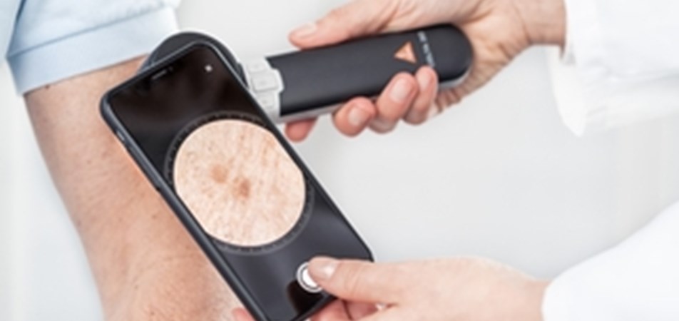
Teledermoscopy uses the camera on an individual’s mobile phone with an attachment that allows the user to take magnified photographs of skin lesions. Dermoscopic-quality pictures of skin lesions can be sent to a specialist for triage. The “store and forward” technology enables provision of dermatologic care services remotely at a later time point for diagnosing and advising management of skin cancer when an in-person clinical visit is not possible.7 Theoretical advantages of teledermoscopy include close inspections of specific skin lesions to improve melanoma early detection for people in underserved or remote areas. Some of the challenges of teledermoscopy include poor image quality and/or lesion selection when photos are taken by patients, inadequate clinical data, and potentially the loss of a local patient–clinician relationship if digital skin assessment is transferred to a metropolitan-based dermatologist.
3D imaging
Technology for total body photography has recently developed from a sequence of separate two-dimensional (2D) images of different parts of the skin’s surface to 3D imaging. The ACEMID VECTRA WB360 3D whole body imaging system (Canfield Scientific, Parsippany, NJ, USA) consists of 92 cameras that simultaneously capture nearly the entire skin surface, including curved surfaces in macro-quality resolution, and then constructs a 3D digital avatar of the individual,8 allowing the precise location of moles across explorations. This image can ensure precise documentation of the exact anatomical location of each lesion to identify changes over time.8 The clinician can review sequential imaging sessions consisting of 3D imaging and determine whether dermoscopy, biopsy or excision is necessary. The advantages of 3D imaging include rapid acquisition of the images,8 software that allows integration of dermoscopy for individual lesions into the 3D avatar, with each lesion given an individual identifier location in the 3D space; and automated assessment of size, colour variation and border irregularity, allowing clinicians to monitor skin lesion stability. The challenge is to enable access to 3D imaging in rural and remote Australia for equitable health service delivery.
Proteomics-based tests for lesion diagnosis
In clinical practice, GPs and dermatologists every so often face the diagnostic dilemma of ambiguous pigmented and non-pigmented lesions.9 Clinicians must balance the risk of leaving a possible melanoma to grow further, with the removal of a benign lesion and related medico-legal, patient and health system concerns, including unwarranted excisions and overdiagnosis.10 Therefore, the ACEMID team is pursuing the idea of a scarless biopsy in the domain of proteomic signatures under an “omics”-based protocol. During a scarless biopsy, the stratum corneum (upper layers of skin) is collected by consecutive application and removal of adhesive discs, applied to the skin’s surface (ie, tape stripping) supported by protein extraction and quantification using mass spectrometry. An advantage of scarless biopsy will be the ability to add to the sensitivity and specificity of existing diagnostic and prognostic markers (unpublished data).
AI-driven algorithms for melanoma diagnosis
AI-enabled medical technologies have the potential to support clinical decision making. The aim of AI is to surpass human cognitive functioning such that automatic decisions can be made autonomously, yet still achieve a reliable diagnostic assessment. Then, sophisticated AI-driven algorithms that evaluate imaging data banks, genomic information, electronic health records, and sociodemographic data can predict diagnosis, prognosis, and optimal management for individuals. For example, the QRISK risk prediction algorithm for future risk of cardiovascular disease uses data from health records,11 and the diabetic retinopathy algorithm uses an imaging data bank of colour fundus photographs.12 These have been approved for clinical use by the National Institute for Health and Care Excellence (QRISK) and the United States Food and Drug Administration (diabetic retinopathy algorithm).
In melanoma diagnosis, AI-driven algorithms reported in laboratory studies have similar diagnostic accuracy relative to that of dermatologists under test conditions.13, 14 Misdiagnosis, overtreatment and under-reporting are current issues in melanoma and skin cancer care. A recent study found that about 55% of melanoma cases (invasive melanoma, 22%) were overdiagnosed10 and associated with iatrogenic harms and increased health care costs. Melanoma misdiagnosis accounts for a greater number of pathology and dermatology malpractice claims compared with other cancers.15 Recent studies report improved accuracy of skin cancer classification by AI-driven approaches versus dermatologists/pathologists using standard clinical practices.16-18 The potential for harm clearly exists in the current standard health care services due to a high rate of false-positive diagnoses, adverse effects, overdiagnosis, and potential overtreatment,19 but as to whether this is improved or worsened with precision medicine technologies remains to be seen.
However, using AI for decision making in the clinical setting needs further study, taking into account the contextual and temporal component of the biologic ecosystem of skin lesions, as well as clinician–AI–patient interactions.18 The ACEMID technology20 proposes automated lesion identification and diagnosis to be used either in a triage capacity before clinical review, whereby lesions suspicious for melanoma would be prioritised, or after clinical review acting as an independent second opinion.21
Evaluation of precision medicine technologies by HTA agencies
Place in the clinical pathway. ACEMID technologies can have multiple places on the clinical pathway. They can be predictive and/or prognostic, may be used as a triage tool for high risk lesions and/or high risk individuals, a diagnostic support tools as either an add-on or a replacement test. They can also be used as one-off or multiple times along the clinical pathway. By providing a baseline, quality assurance becomes possible.
Uncertainty. Multilevel uncertainty in clinical and economic models is likely to arise from wide precision estimates of effect due to very small sample sizes of like-individuals. ACEMID technologies may stratify people into small subgroups based on imaging, genetic or protein signatures. Uncertainty in estimates of effectiveness may also result from the dynamic nature of the technologies that are likely to improve in their precision and relevance over time. For example, AI-driven algorithms are continually building upon new image datasets to improve their diagnostic sensitivity and specificity. Applications with frequently updated interfaces and changeable hosting platforms (eg, those providing automated skin lesion assessment or clinician-assessed teledermoscopy) also represent technologies where small, yet frequent changes may alter the uncertainty around the technology’s efficacy, effectiveness or cost-effectiveness.
Equity. The disproportionate use of new precision medicine technologies for individuals with higher income, compared with individuals with lower socio-economic level, may widen the current gap in melanoma health outcomes. Although we have seen a growth in digital health care throughout the coronavirus disease 2019 (COVID-19) pandemic, many patients could be excluded from these health technology-driven services if disproportionate access continues to be related to socio-economic status. Interconnected with this concern is a bias that many algorithms have so far excluded skin of colour and uncommon presentations of melanoma and other rare malignant skin neoplasms in their datasets and image banks.22
Shelf-life of the guidance. Finally, the duration of time in which the effectiveness evidence remains current for precision medicine diagnostics may be short before they are replaced with the next iteration or model. For example, other systems are in development that will provide total body images with dermoscopy quality in the future.23 As precision medicine technologies are increasingly used, the methods and processes of HTA will need to adapt to maintain their objective of evaluating whether a health technology investment is good value for money.
Recommendations
Conclusion
Precision medicine technologies have the potential to revolutionise the way melanoma is detected and managed. ACEMID focuses on 3D imaging and telemedicine research, in which Australia will lead the evidence generation of diagnostic efficacy, and real-world implementation into primary and specialist care. To assess the value of these precision medicine technologies, HTA agencies will need to accommodate evidence from different study designs, dynamic modelling from evolving biomarkers and algorithms, a mechanism for assessing the impact on health equity, and rapid updates as technology changes.
Acknowledgements:
Rachael Morton is supported by a National Health and Medical Research Council (NHMRC) Emerging Leadership-2 Fellowship (1194703). We acknowledge the Australian Cancer Research Foundation for financial support to the ACEMID program. Victoria Mar received the NHMRC Clinical Trials and Cohort Studies Grant (2001517), and Monika Janda received the NHMRC Centre of Research Excellence (2006551) and the NHMRC Synergy Grant (2009923). We thank the following ACEMID Team members for their contribution to this manuscript: Anne Cust (Daffodil Centre, University of Sydney and Cancer Council NSW), Joanne Aitken (Cancer Council Queensland); Richard Scolyer (Melanoma Institute Australia, University of Sydney), Pascale Guitera (Sydney Melanoma Diagnostic Centre, Royal Prince Alfred Hospital), Liam Caffery (Centre for Online Health, University of Queensland), Rory Wolfe (Monash University), and Graham Mann (John Curtin School of Medical Research, Australian National University).
Open access:
Open access publishing facilitated by The University of Sydney, as part of the Wiley - The University of Sydney agreement via the Council of Australian University Librarians.
Competing interests:
H Peter Soyer is a shareholder of MoleMap NZ and E-Derm-Consult and undertakes regular teledermatological reporting for both companies; is a medical consultant for Canfield Scientific, MoleMap Australia, Blaze Bioscience and Revenio Research; and is a medical advisor for First Derm.
Provenance:
Not commissioned; externally peer reviewed.