To recover your password please fill in your email address
Please fill in below form to create an account with us

Benefits and harms of CT and PET/CT surveillance imaging for melanoma.
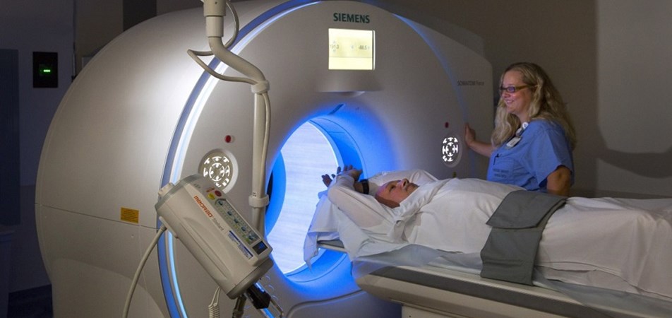
The use of computed tomography (CT) or positron emission tomography (PET)/CT surveillance imaging in post-melanoma treatment follow-up schedules is increasing rapidly. However good estimates of the radiation-attributable cancer risk associated with long-term surveillance imaging is lacking. The aim of this retrospective study is to quantify excess lifetime risk of new cancers attributable to CT or PET/CT surveillance imaging. A Monte Carlo simulation model is used to calculate life-long radiation exposure. Imaging protocols included FDG-PET and CT chest-abdomen-pelvis and CT brain imaging at intervals of three, six or 12-months, over a three, five or 10-year surveillance duration. The model represents imaging practices common to leading tertiary melanoma treatment centres in the United States, Europe and Australia. Simulation model participants include asymptomatic adults treated for AJCC stage IIIA-D melanoma aged 20, 40, or 60-years at diagnosis.
|
Collaborators: |
Columbia University, Melanoma Institute Australia, University of Sydney, St Vincent’s Hospital, Royal Prince Alfred Hospital |
|
Funded by: |
Cancer Australia, Priority-driven Collaborative Cancer Research Scheme grant #1129568 |
|
Chief investigators: |
Rachael L Morton, Anna Nording, Andrew Einstein, John F Thompson, Louise Emmett, Robyn Saw, Omgo Nieweg |
|
Contact: |